About Thermography
What is Thermography?

Breast Thermography is an imaging procedure where infrared images of the breast are analyzed and rated to determine breast health. Thermography is non-invasive, safe and painless – no radiation or compression is used.
Breast Thermography is based on the premise that before the growth of abnormal cells is possible, a constant blood supply must be circulated to the growth area. Chemical and blood vessel activity in both pre-cancerous tissue and the area surrounding a developing breast cancer is almost always higher than in the normal breast. Since pre-cancerous and cancerous masses are highly metabolic tissues, they need an abundant supply of nutrients to maintain their growth and this can increase the surface temperatures of the breast. Breast Thermography measures the heat generated by the microcirculation of blood in the breast during this process.
Why Thermography?
Thermal imaging has been an available medical test for more than 50 years now. Thermography is a totally safe, does not expose a patient to any form of radiation, it’s non-invasive, painless test with absolutely no adverse effects and with no contraindications. It can be used at any age and provides a diagnostic tool far superior to others in early the stages of some diseases.
It is always important to stay proactive and well informed in your own health care. In addition to your regular medical breast examinations, consider regular breast screening with Thermography, the results could change your life. Thermography is one of the best early warning systems available today.
How is it Done?
A state-of-the-art FDA approved infrared imaging camera measures skin temperature and transmits this information to a computer which converts these radiometric measurements into images. These thermographic findings are evaluated based on 32 standard thermological signs and quantification criteria as set forth and accepted standards of clinical thermology. Temperature measurements will differ in various parts of the body, but in a normal healthy individual, the temperature changes should be relatively symmetrical. Once the images are acquired in a controlled setting they are sent for processing to our board certified clinical Thermologists for analysis and interpretation. Your written report is then returned to your imaging center in a few days and presented back to you with an explanation and follow up recommendations. Studies show that when breast thermography is used as part of a multimodal approach (clinical examination + thermography + mammography), 95% of early stage cancers can be detected.
Symmetrical distribution in both breasts
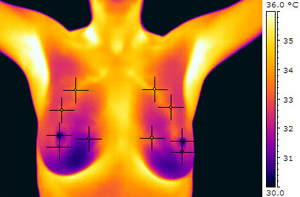
Normal breast – Low Risk.
Note very high temperatures and
asymmetrical presentation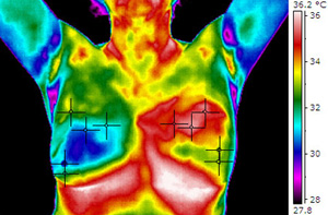
Cancer of the Left Breast.
